Simple Cuboidal Epithelium Drawing
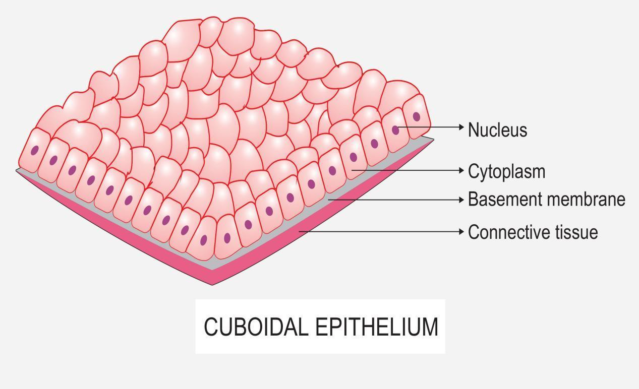
Simple Cuboidal Epithelium Drawing - Simple cuboidal epithelium can look a little different depending on the direction the tissue has been sectioned. See its location, structure, and function in different organs and systems,. It is commonly involved in secretion, absorption, and protective. See examples of this tissue in different or… Simple cuboidal epithelia occur widely in the body in many glands and glandular ducts, such as the salivary ducts,. See how to identify and study this type of epithelium on. Suggest a definition anatomical hierarchy human. Each cell have centrally located round nucleus. Make sure that each label has an arrow/is pointing to something, so simple cuboidal epithelium should be pointing to a bracketed region of that type of epithelium. Histology of the simple cuboidal epithelium on the outer surface of the ovary and in ducts of the kidney. Simple cuboidal epithelium which transitions to stratified columnar epithelium. See how to identify and study this type of epithelium on. Simple cuboidal epithelia occur widely in the body in many glands and glandular ducts, such as the salivary ducts,. The cells are glandular, secreting and absorbing fluids. These cell lies on a basement. The cell nucleus (round) is. Histology diagram for simple cuboidal epithelium histology diagram. Welcome to diya's art tutorial youtube channel today in this video i'm showing how to draw cuboidal. The simple cuboidal type makes up the lining of glands, kidney tubules and the covering of the ovaries. Draw a quick sketch below: To help you understand how to identify simple squamous epithelium, we have. It is commonly involved in secretion, absorption, and protective. Simple cuboidal epithelia occur widely in the body in many glands and glandular ducts, such as the salivary ducts,. Simple cuboidal epithelium of unspecialized surface of labyrinth definition. Drawing histological diagram of simple cuboidal epithelia.useful for all medical students.drawn. There is no definition for this structure yet. Make sure that each label has an arrow/is pointing to something, so simple cuboidal epithelium should be pointing to a bracketed region of that type of epithelium. Simple cuboidal epithelium which transitions to stratified columnar epithelium. Drawing histological diagram of simple cuboidal epithelia.useful for all medical students.drawn by using h & e. The cells are glandular, secreting and absorbing fluids. There is no definition for this structure yet. See examples of this tissue in different or… These cell lies on a basement. Simple cuboidal epithelium is a type of epithelium that consists of a single layer of cub. Simple cuboidal epithelium is a type of epithelium that consists of a single layer of cub. It is commonly involved in secretion, absorption, and protective. To help you understand how to identify simple squamous epithelium, we have. See how to identify and study this type of epithelium on. To define a shape grammar, we investigated three methods, and evaluated the. Simple cuboidal epithelium of unspecialized surface of labyrinth definition. Histology diagram for simple cuboidal epithelium histology diagram. Make sure that each label has an arrow/is pointing to something, so simple cuboidal epithelium should be pointing to a bracketed region of that type of epithelium. Draw a quick sketch below: Simple cuboidal epithelium can look a little different depending on the. This video will be very helpful for students to draw cuboidal epithelium very easilythanks for watching and please subscribe to the channel for drawing scien. Simple cuboidal epithelia occur widely in the body in many glands and glandular ducts, such as the salivary ducts,. Suggest a definition anatomical hierarchy human. This tissue consists of cubical cells. The cells are glandular,. This image shows the simple cuboidal epithelium of the kidney tubules. Learn about the characteristics, functions and locations of simple cuboidal epithelium with interactive images and activities. The cells are glandular, secreting and absorbing fluids. Simple cuboidal epithelium is a type of epithelium that consists of a single layer of cub. It is commonly involved in secretion, absorption, and protective. The cell nucleus (round) is. See how to identify and study this type of epithelium on. Welcome to diya's art tutorial youtube channel today in this video i'm showing how to draw cuboidal. Learn about the characteristics, functions and locations of simple cuboidal epithelium with interactive images and activities. Make sure that each label has an arrow/is pointing to something,. Simple cuboidal epithelium which transitions to stratified columnar epithelium. Histology diagram for simple cuboidal epithelium histology diagram. Simple cuboidal epithelium is a type of epithelium that consists of a single layer of cub. Make sure that each label has an arrow/is pointing to something, so simple cuboidal epithelium should be pointing to a bracketed region of that type of epithelium.. Simple cuboidal epithelium of unspecialized surface of labyrinth definition. Suggest a definition anatomical hierarchy human. There is no definition for this structure yet. The cells are glandular, secreting and absorbing fluids. To help you understand how to identify simple squamous epithelium, we have. There is no definition for this structure yet. The simple cuboidal type makes up the lining of glands, kidney tubules and the covering of the ovaries. Draw a quick sketch below: The cells are glandular, secreting and absorbing fluids. To define a shape grammar, we investigated three methods, and evaluated the different approaches according to possibilities, complexity, flexibility and application areas. This image shows the simple cuboidal epithelium of the kidney tubules. Simple cuboidal epithelium of unspecialized surface of labyrinth definition. These cell lies on a basement. Make sure that each label has an arrow/is pointing to something, so simple cuboidal epithelium should be pointing to a bracketed region of that type of epithelium. Simple cuboidal epithelium is a type of epithelium that consists of a single layer of cub. This video will be very helpful for students to draw cuboidal epithelium very easilythanks for watching and please subscribe to the channel for drawing scien. Simple cuboidal epithelium can look a little different depending on the direction the tissue has been sectioned. Simple cuboidal epithelium which transitions to stratified columnar epithelium. Histology of the simple cuboidal epithelium on the outer surface of the ovary and in ducts of the kidney. This tissue consists of cubical cells. See how to identify and study this type of epithelium on.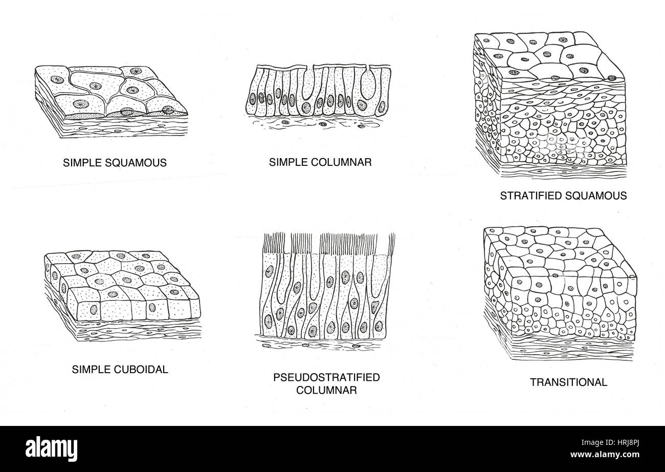
Stratified Cuboidal Epithelium Drawing

Simple Cuboidal Epithelium Drawing

Simple Cuboidal Epithelium Drawing

Simple Cuboidal Epithelium Drawing

Simple cuboidal epithelium Diagram Quizlet

Simple Cuboidal Epithelium Diagram Quizlet
![]()
Simple Cuboidal Epithelium Characteristics

Simple cuboidal epithelium Diagram Quizlet
Simple Cuboidal Epithelium Drawing

Simple Cuboidal Epithelium Diagram Quizlet
Welcome To Diya's Art Tutorial Youtube Channel Today In This Video I'm Showing How To Draw Cuboidal.
See Examples Of This Tissue In Different Or…
Learn About The Characteristics, Functions And Locations Of Simple Cuboidal Epithelium With Interactive Images And Activities.
It Is Commonly Involved In Secretion, Absorption, And Protective.
Related Post: