Salivary Glands Drawing
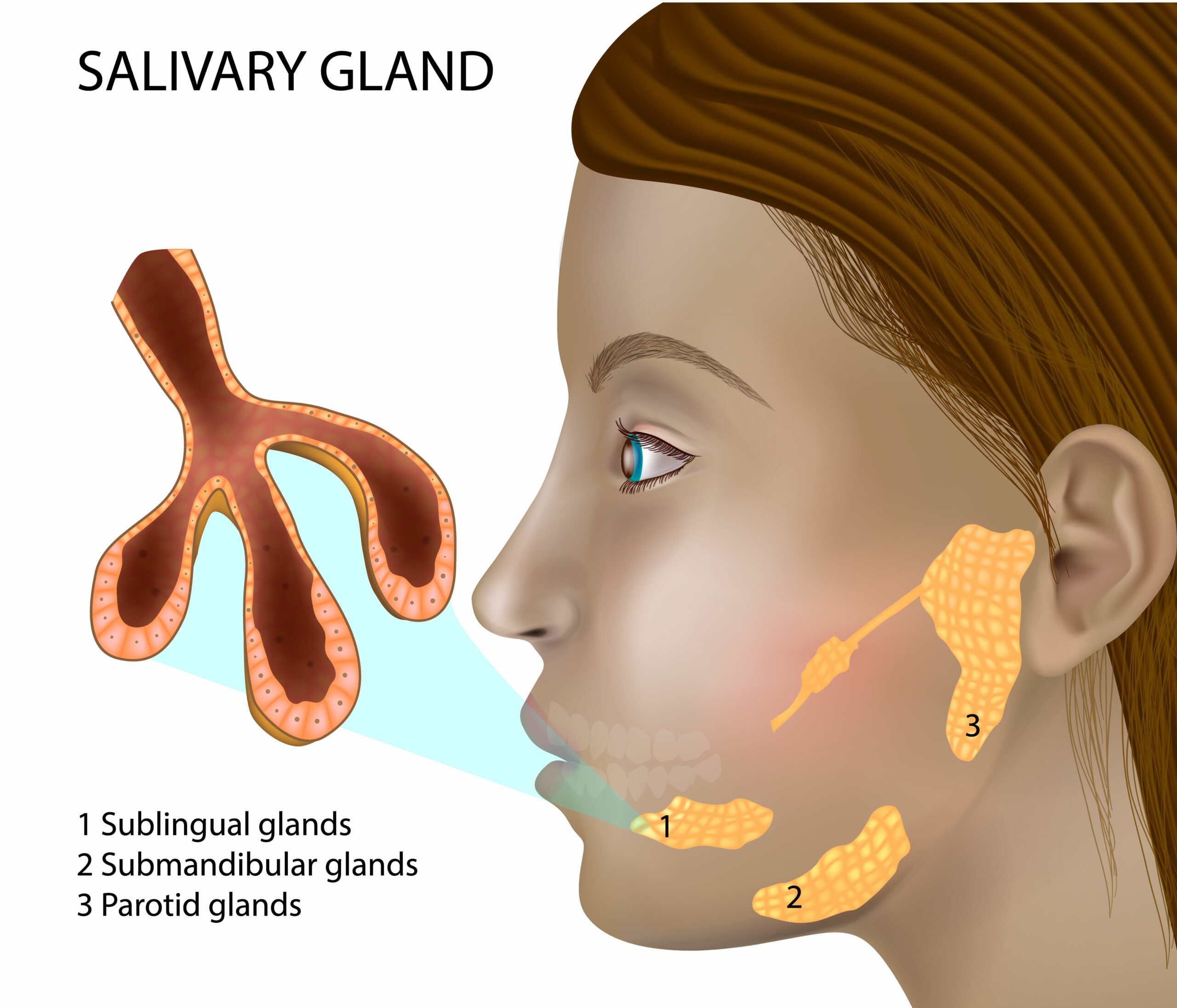
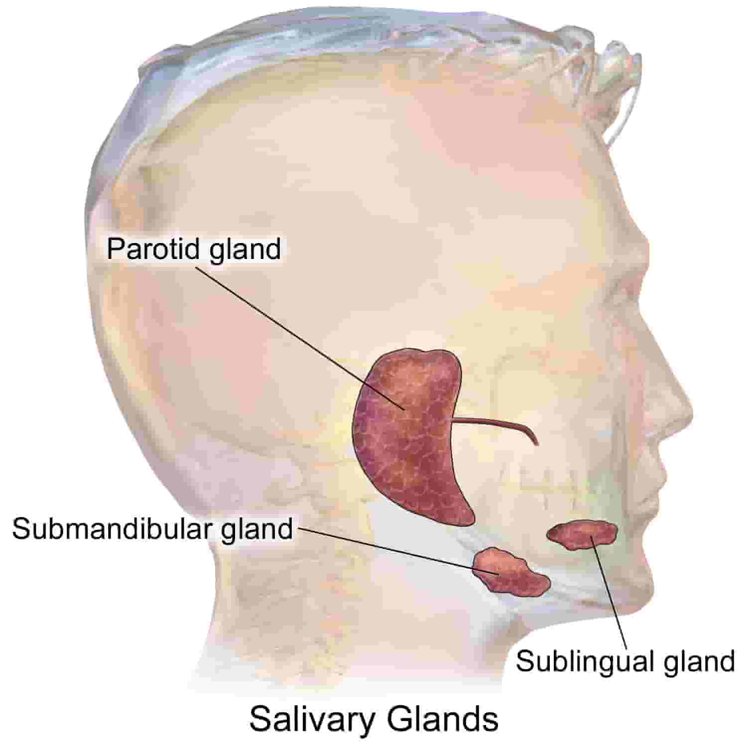
Salivary Glands Drawing - Location of salivary glands and ducts. Saliva is produced by three pairs of major and minor salivary glands. Neatly labeled and easy to draw, this. Diagram showing inside of mouth and salivary glands. Saliva aids in moistening food, digestion, lubrication, and oral hygiene. These salivary glands secrete saliva into the mouth via the various ducts. Anatomy of the salivary glands; Download scientific diagram | anatomy of the salivary glands. Find & download free graphic resources for salivary glands vectors, stock photos & psd files. Drawing shows a cross section of the head and the three main pairs of salivary glands. For similar images and pathology, see smart.servier.com. In this article we will discuss about the structure of salivary glands of human with its suitable diagram. Find & download free graphic resources for salivary glands vectors, stock photos & psd files. The major salivary glands, namely the parotid, submandibular, and sublingual glands contribute approximately. Common salivary glands such as the parotid and submandibular glands can be adequately visualized using current clinical imaging modalities such as computed tomography (ct),. These salivary glands secrete saliva into the mouth via the various ducts. These salivary glands secrete saliva into the mouth via the various ducts. There are two types of acini in the. The parotid glands are in front of and just below each ear; Retrieved from anatomy & physiology by open learning. Saliva aids in moistening food, digestion, lubrication, and oral hygiene. Salivary glands are exocrine glands responsible for producing and secreting saliva into the oral cavity. Neatly labeled and easy to draw, this. These glands secrete saliva in the oral cavity through a system of ducts. They are responsible for producing saliva helping in digestion and oral health. Submandibular gland, parotid gland, facial muscles, hemisected tongue, submental triangle, common carotid artery, carotid bifurcation, vagus. Salivary gland diagram shows its structure in the mouth. Anatomy of the salivary glands; The major salivary glands, namely the parotid, submandibular, and sublingual glands contribute approximately. Browse 121 saliva gland photos and images available, or start a new search to explore more photos. Submandibular gland, parotid gland, facial muscles, hemisected tongue, submental triangle, common carotid artery, carotid bifurcation, vagus. Secretory unit of salivary gland is an acinus. The parotid glands are in front of and just below each ear; Anatomy of the salivary glands; Retrieved from anatomy & physiology by open learning. Submandibular gland, parotid gland, facial muscles, hemisected tongue, submental triangle, common carotid artery, carotid bifurcation, vagus. The salivary gland diagram provides a clear visual representation of the major salivary glands: Download scientific diagram | anatomy of the salivary glands. They are responsible for producing saliva helping in digestion and oral health. The major salivary glands, namely the parotid, submandibular, and. Find & download free graphic resources for salivary glands vectors, stock photos & psd files. Anatomy of the salivary glands; Secretory unit of salivary gland is an acinus. Free for commercial use high quality images Anatomy of the salivary glands. The gastrointestinal tract the appendix. Salivary glands are exocrine glands responsible for producing and secreting saliva into the oral cavity. Anatomy of the salivary glands; Anatomy of the salivary glands. Secretory unit of salivary gland is an acinus. Common salivary glands such as the parotid and submandibular glands can be adequately visualized using current clinical imaging modalities such as computed tomography (ct),. Drawing shows a cross section of the head and the three main pairs of salivary glands. Anatomy of the salivary glands; The salivary gland diagram provides a clear visual representation of the major salivary glands: The. Find & download free graphic resources for salivary glands vectors, stock photos & psd files. The diagram of salivary glands illustrates. Anatomy of the salivary glands. The parotid glands are in front of and just below each ear; These glands secrete saliva in the oral cavity through a system of ducts. Retrieved from anatomy & physiology by open learning. Neatly labeled and easy to draw, this. Submandibular gland, parotid gland, facial muscles, hemisected tongue, submental triangle, common carotid artery, carotid bifurcation, vagus. The parotid glands are in front of and just below each ear; The major salivary glands, namely the parotid, submandibular, and sublingual glands contribute approximately. Diagram showing inside of mouth and salivary glands. Saliva aids in moistening food, digestion, lubrication, and oral hygiene. A axial drawing depicts the parotid space and its relationship with surrounding neck spaces. Retrieved from anatomy & physiology by open learning. Common salivary glands such as the parotid and submandibular glands can be adequately visualized using current clinical imaging modalities such. Common salivary glands such as the parotid and submandibular glands can be adequately visualized using current clinical imaging modalities such as computed tomography (ct),. There are two types of acini in the. In this article we will discuss about the structure of salivary glands of human with its suitable diagram. Drawing shows a cross section of the head and the three main pairs of salivary glands. Anatomy of the salivary glands; These glands secrete saliva in the oral cavity through a system of ducts. They are responsible for producing saliva helping in digestion and oral health. Anatomy of the salivary glands. Saliva aids in moistening food, digestion, lubrication, and oral hygiene. The gastrointestinal tract the appendix. A axial drawing depicts the parotid space and its relationship with surrounding neck spaces. Salivary gland diagram shows its structure in the mouth. The parotid glands are in front of and just below each ear; The major salivary glands, namely the parotid, submandibular, and sublingual glands contribute approximately. Find & download free graphic resources for salivary glands vectors, stock photos & psd files. The salivary gland diagram provides a clear visual representation of the major salivary glands:The mouth, throat, nose and ears Macmillan Cancer Support

Physiology Glossary Salivary Gland Physiology Draw It to Know It
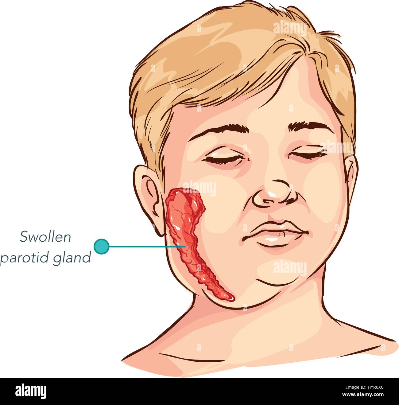
children salivary gland swelling vector illustration Stock Vector Image

Salivary glands hires stock photography and images Alamy

Sublingual Salivary Gland. Vector Illustration on Isolated Background
Salivary Glands Richmond ENT
Salivary glands ClipArt ETC
Salivary gland function and structure Anatomy And Physiology

Digestion in the Mouth MooMooMath and Science

Salivary glands, illustration Stock Image C050/6261 Science Photo
These Salivary Glands Secrete Saliva Into The Mouth Via The Various Ducts.
Free For Commercial Use High Quality Images
Location Of Salivary Glands And Ducts.
Anatomy Of The Salivary Glands;
Related Post: