Plasma Membrane Drawing Labeled
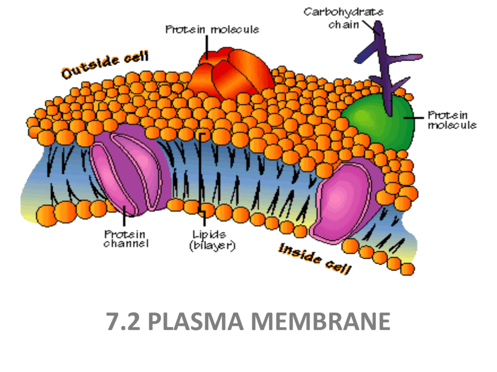
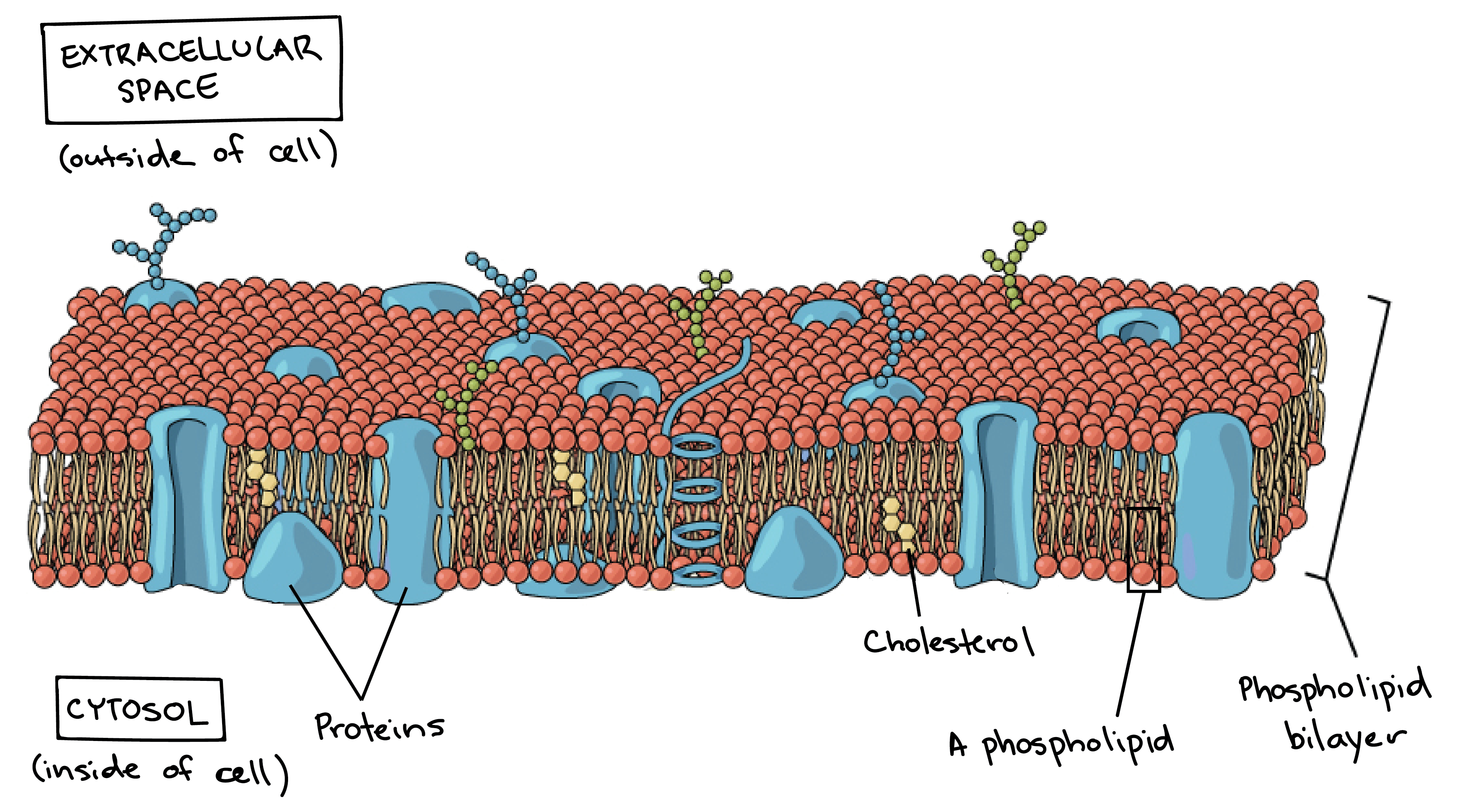
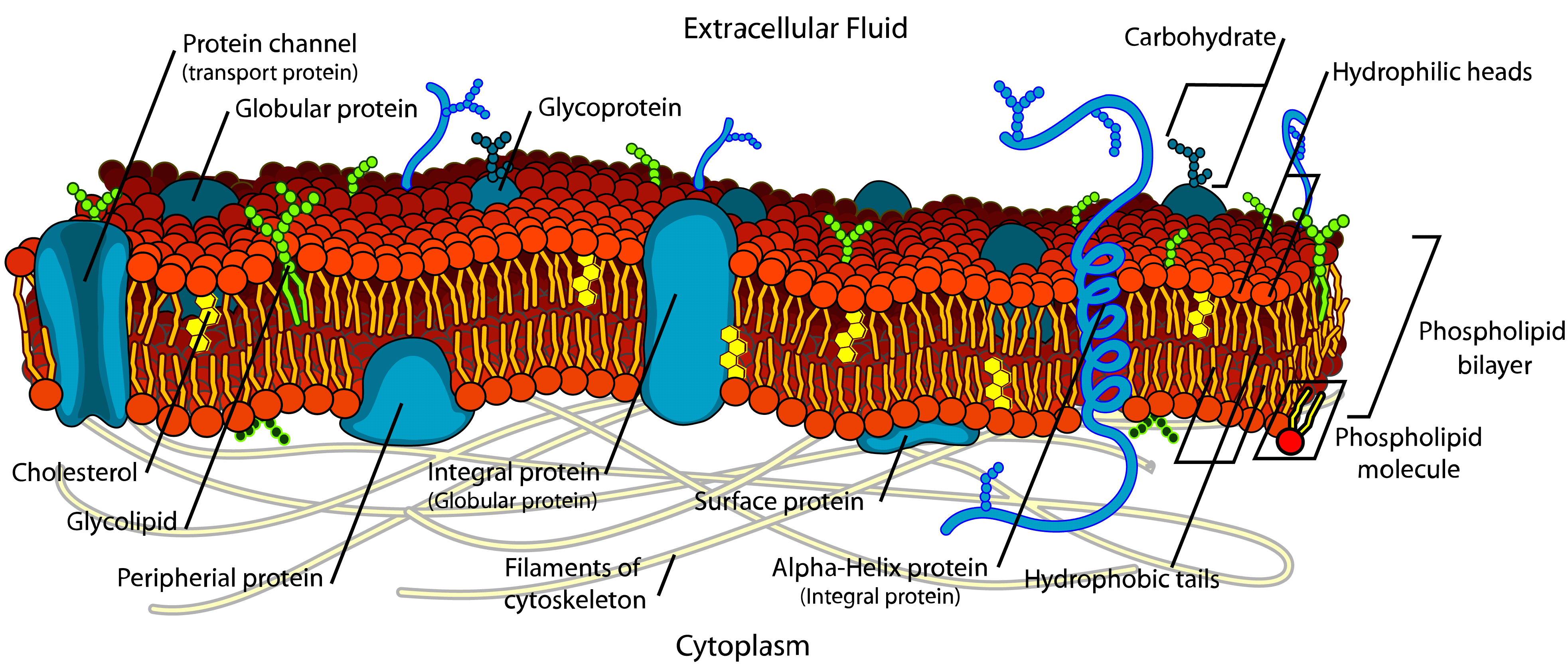
Plasma Membrane Drawing Labeled - The cell membrane, also known as the plasma membrane, is a double layer of lipids and proteins that surrounds a cell. In eukaryotic cells, the plasma membrane surrounds a cytoplasm filled with ribosomes and organelles. When drawing and labeling a diagram of the plasma membrane you should be sure to include: Let's begin with the structure of the plasma membrane, whose key components are phospholipids and proteins. This theory, compared to earlier theories, best explains both microscopic observations and the. Organelles are structures that are themselves encased in membranes. Phospholipids form a bilayer in which their nonpolar, hydrophobic tails are. It separates the cytoplasm (the contents of the cell). Nicolson proposed a new model of the plasma membrane. The plasma membrane is a protective barrier that surrounds cells. Plasma membrane is a fluid mosaic of proteins, lipids and carbohydrates. This theory, compared to earlier theories, best explains both microscopic observations and the. The plasma membrane picture provided above shows the detailed structure of the plasma membrane. A plasma membrane’s principal components are lipids (phospholipids and cholesterol), proteins, and carbohydrates attached to some of the lipids and proteins. The plasma membrane is a protective barrier that surrounds cells. It separates the cytoplasm (the contents of the cell). The phospholipid bilayer with hydrophobic 'tails' and hydrophilic 'heads' of the. In eukaryotic cells, the plasma membrane surrounds a cytoplasm filled with ribosomes and organelles. The fluid mosaic model describes the structure of the plasma membrane as a mosaic of components —including phospholipids, cholesterol, proteins, and. 2.4.2 explain how the hydrophobic and hydrophilic properties of. This theory, compared to earlier theories, best explains both microscopic observations and the. The plasma membrane is a protective barrier that surrounds cells. Both prokaryotic and eukaryotic cells have plasma membranes, but they vary among different organisms. Organelles are structures that are themselves encased in membranes. 2.4.1 draw and label a diagram to show the structure of membranes. Phospholipids form a bilayer in which their nonpolar, hydrophobic tails are. Nicolson proposed a new model of the plasma membrane. The fluid mosaic model describes the structure of the plasma membrane as a mosaic of components —including phospholipids, cholesterol, proteins, and. The phospholipid bilayer with hydrophobic 'tails' and hydrophilic 'heads' of the. Organelles are structures that are themselves encased in. Phospholipids form a bilayer in which their nonpolar, hydrophobic tails are. When drawing and labeling a diagram of the plasma membrane you should be sure to include: Plasma membrane is a fluid mosaic of proteins, lipids and carbohydrates. The plasma membrane picture provided above shows the detailed structure of the plasma membrane. Let's begin with the structure of the plasma. 2.4.2 explain how the hydrophobic and hydrophilic properties of. It separates the cytoplasm (the contents of the cell). 2.4.1 draw and label a diagram to show the structure of membranes. Just as the outer layer of your skin separates your body from its environment, the cell membrane (also known as the plasma membrane) separates the inner contents of a cell. Phospholipids form a bilayer in which their nonpolar, hydrophobic tails are. The phospholipid bilayer with hydrophobic 'tails' and hydrophilic 'heads' of the. This theory, compared to earlier theories, best explains both microscopic observations and the. Organelles are structures that are themselves encased in membranes. The fluid mosaic model describes the structure of the plasma membrane as a mosaic of components. A plasma membrane’s principal components are lipids (phospholipids and cholesterol), proteins, and carbohydrates attached to some of the lipids and proteins. Plasma membrane is a fluid mosaic of proteins, lipids and carbohydrates. When drawing and labeling a diagram of the plasma membrane you should be sure to include: Both prokaryotic and eukaryotic cells have plasma membranes, but they vary among. This theory, compared to earlier theories, best explains both microscopic observations and the. The plasma membrane picture provided above shows the detailed structure of the plasma membrane. Both prokaryotic and eukaryotic cells have plasma membranes, but they vary among different organisms. It separates the cytoplasm (the contents of the cell). Nicolson proposed a new model of the plasma membrane. When drawing and labeling a diagram of the plasma membrane you should be sure to include: Let's begin with the structure of the plasma membrane, whose key components are phospholipids and proteins. It separates the cytoplasm (the contents of the cell). This theory, compared to earlier theories, best explains both microscopic observations and the. 2.4.2 explain how the hydrophobic and. The cell membrane, also known as the plasma membrane, is a double layer of lipids and proteins that surrounds a cell. A plasma membrane’s principal components are lipids (phospholipids and cholesterol), proteins, and carbohydrates attached to some of the lipids and proteins. A phospholipid is a molecule. Plasma membrane is a fluid mosaic of proteins, lipids and carbohydrates. The fluid. The phospholipid bilayer with hydrophobic 'tails' and hydrophilic 'heads' of the. This theory, compared to earlier theories, best explains both microscopic observations and the. The plasma membrane is a protective barrier that surrounds cells. Phospholipids form a bilayer in which their nonpolar, hydrophobic tails are. When drawing and labeling a diagram of the plasma membrane you should be sure to. It separates the cytoplasm (the contents of the cell). 2.4.1 draw and label a diagram to show the structure of membranes. A phospholipid is a molecule. In eukaryotic cells, the plasma membrane surrounds a cytoplasm filled with ribosomes and organelles. Plasma membrane is a fluid mosaic of proteins, lipids and carbohydrates. When drawing and labeling a diagram of the plasma membrane you should be sure to include: Nicolson proposed a new model of the plasma membrane. Both prokaryotic and eukaryotic cells have plasma membranes, but they vary among different organisms. A plasma membrane’s principal components are lipids (phospholipids and cholesterol), proteins, and carbohydrates attached to some of the lipids and proteins. 2.4.2 explain how the hydrophobic and hydrophilic properties of. The plasma membrane picture provided above shows the detailed structure of the plasma membrane. The cell membrane, also known as the plasma membrane, is a double layer of lipids and proteins that surrounds a cell. The fluid mosaic model describes the structure of the plasma membrane as a mosaic of components —including phospholipids, cholesterol, proteins, and. This theory, compared to earlier theories, best explains both microscopic observations and the. Phospholipids form a bilayer in which their nonpolar, hydrophobic tails are. Organelles are structures that are themselves encased in membranes.
Labeled Diagram Of Plasma Membrane Best Of Plasma Membrane Diagrams

Main Component of Plasma Membrane

Biological Diagram Show Structure Of Cell Membrane Or Plasma Membrane

Realistic human cell anatomy infographics with diagram showing plasma

Plasma Membrane Diagrams 101 Diagrams
STRUCTURE of PLASMA MEMBRANE
Labeled Diagram Of The Plasma Membrane

5.4 Plasma Membrane Biology LibreTexts
Plasma Membrane Labelled Diagram

Functions of the Plasma Membrane Biology Wise
Let's Begin With The Structure Of The Plasma Membrane, Whose Key Components Are Phospholipids And Proteins.
The Phospholipid Bilayer With Hydrophobic 'Tails' And Hydrophilic 'Heads' Of The.
The Plasma Membrane Is A Protective Barrier That Surrounds Cells.
Just As The Outer Layer Of Your Skin Separates Your Body From Its Environment, The Cell Membrane (Also Known As The Plasma Membrane) Separates The Inner Contents Of A Cell From Its Exterior.
Related Post: