Knee Joint Drawing
Knee Joint Drawing - Learn about the structure and function of the knee joint with diagrams and examples. To add detail to the knee joint, start by drawing the condyles of the femur and the tibia. This image shows the knee joint and patella from ventral, medial and dorsal. Free for commercial use high quality images See diagrams and pictures of the bones, ligaments, cartilage, and muscles of the knee. One between the femur and tibia and one between the femur and patella. The femur, tibia and patella. It's a hinge joint that allows the legs to move and bend, and is. The joint surfaces are lined with hyaline cartilage and are enclosed within a single joint cavity. Knee anatomy involves different structures in the knee joint and how they connect to each other. The tibiofemoral joint and patellofemoral joint. Find the perfect knee joint drawing stock photo, image, vector, illustration or 360 image. It is a complex hinge joint composed of two articulations; In this joint, the flat surface of the shinbone rotates around the cylindrical surface of the thighbone. Free for commercial use high quality images Find out how to create a knee anatomy diagram by hand or online using edrawmax tool. It can be subdivided into bones, cartilages, ligaments, tendons and muscles. One between the femur and tibia and one between the femur and patella. Anatomy of the knee joint. Knee anatomy involves different structures in the knee joint and how they connect to each other. Sagittal view of the knee joint showing the patellofemoral and tibiofemoral joints. In this video lesson, you will discover how to draw knees from life with the necessary knowledge of this joint's proportions and anatomy. In this joint, the flat surface of the shinbone rotates around the cylindrical surface of the thighbone. Let us examine the knee anatomy. See the. Anterior, medial and posterior view. See diagrams and pictures of the bones, ligaments, cartilage, and muscles of the knee. These are the bumpy parts of the bone that fit together to form the knee joint. Knee anatomy involves different structures in the knee joint and how they connect to each other. It's a hinge joint that allows the legs to. Learn about the structure and function of the knee joint with diagrams and examples. Free for commercial use high quality images When drawing a knee, we have two big volumes—the. To add detail to the knee joint, start by drawing the condyles of the femur and the tibia. The knee joint is a synovial joint that connects three bones; One between the femur and tibia and one between the femur and patella. The femur, tibia and patella. 25,000+ vectors, stock photos & psd files. Find & download free graphic resources for knee joint drawing. In this joint, the flat surface of the shinbone rotates around the cylindrical surface of the thighbone. These are the bumpy parts of the bone that fit together to form the knee joint. In this video lesson, you will discover how to draw knees from life with the necessary knowledge of this joint's proportions and anatomy. Sagittal view of the knee joint showing the patellofemoral and tibiofemoral joints. See diagrams and pictures of the bones, ligaments, cartilage,. This image shows the knee joint and patella from ventral, medial and dorsal. In this joint, the flat surface of the shinbone rotates around the cylindrical surface of the thighbone. It can be subdivided into bones, cartilages, ligaments, tendons and muscles. Find the perfect knee joint drawing stock photo, image, vector, illustration or 360 image. To add detail to the. Find the perfect knee joint drawing stock photo, image, vector, illustration or 360 image. It can be subdivided into bones, cartilages, ligaments, tendons and muscles. In this video lesson, you will discover how to draw knees from life with the necessary knowledge of this joint's proportions and anatomy. The knee joint is the largest joint in the body and connects. It is a complex hinge joint composed of two articulations; In this joint, the flat surface of the shinbone rotates around the cylindrical surface of the thighbone. See the pictures and anatomy description of knee joint bones, cartilage, ligaments, muscle and tendons with resources for knee problems & injuries. The knee joint is the largest joint in the body and. The knee joint is a synovial joint that connects three bones; See the pictures and anatomy description of knee joint bones, cartilage, ligaments, muscle and tendons with resources for knee problems & injuries. Knee anatomy involves different structures in the knee joint and how they connect to each other. When drawing a knee, we have two big volumes—the. Learn about. In this joint, the flat surface of the shinbone rotates around the cylindrical surface of the thighbone. The knee joint joins the thigh with the leg and consists of two articulations: 25,000+ vectors, stock photos & psd files. Knee anatomy involves different structures in the knee joint and how they connect to each other. Sagittal view of the knee joint. It can be subdivided into bones, cartilages, ligaments, tendons and muscles. The tibiofemoral joint and patellofemoral joint. To add detail to the knee joint, start by drawing the condyles of the femur and the tibia. The knee joint is a synovial joint that connects three bones; Sagittal view of the knee joint showing the patellofemoral and tibiofemoral joints. The main function of this. The femur, tibia and patella. When drawing a knee, we have two big volumes—the. See the pictures and anatomy description of knee joint bones, cartilage, ligaments, muscle and tendons with resources for knee problems & injuries. Anterior, medial and posterior view. It's a hinge joint that allows the legs to move and bend, and is. Knee anatomy involves different structures in the knee joint and how they connect to each other. Let us examine the knee anatomy. One between the femur and tibia and one between the femur and patella. 25,000+ vectors, stock photos & psd files. Find out how to create a knee anatomy diagram by hand or online using edrawmax tool.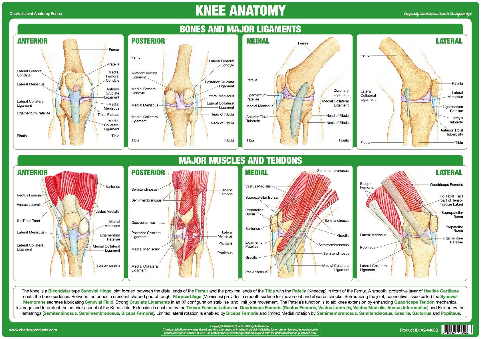
Knee Joint Anatomy Poster
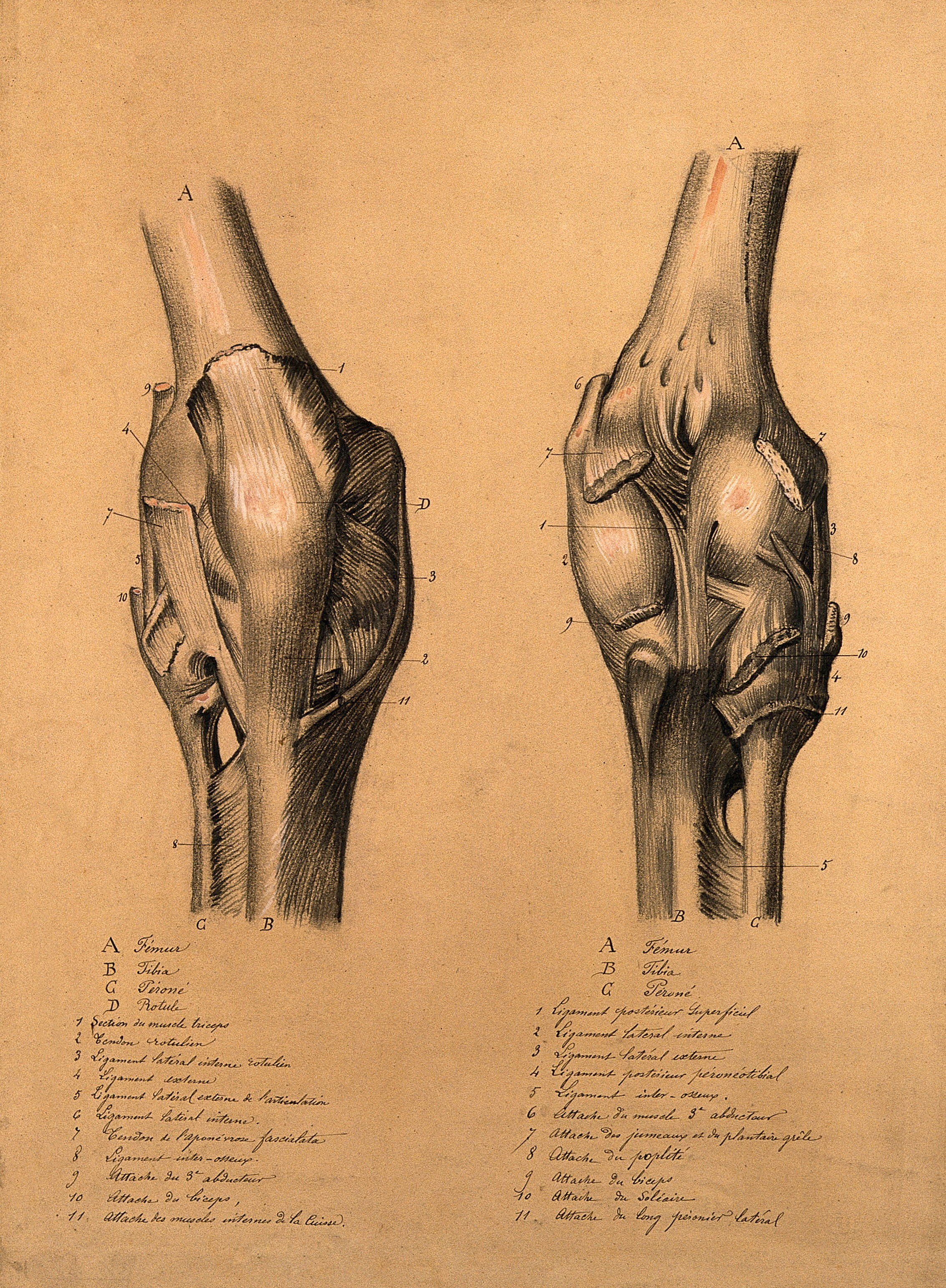
The knee joint two studies, showing the bones and ligaments of the

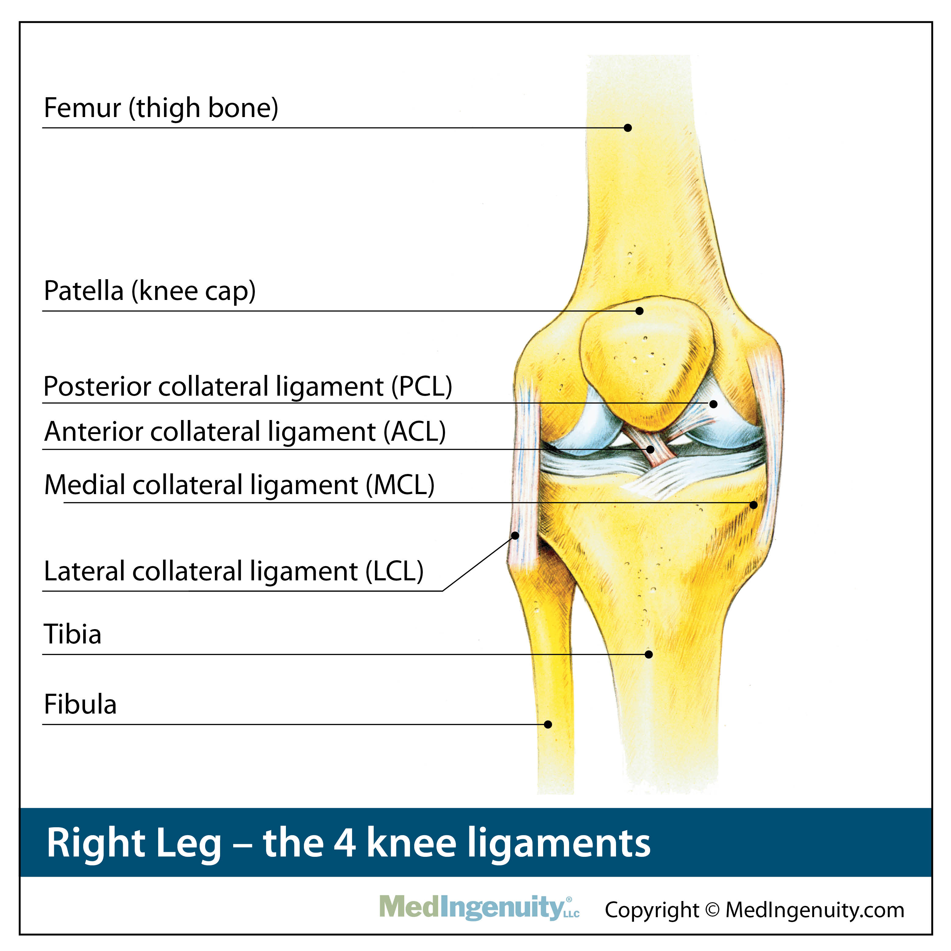
Knee Ligaments JOI Jacksonville Orthopaedic Institute
Orthopedic Anatomy Library Northwest Hills Surgical Hospital in
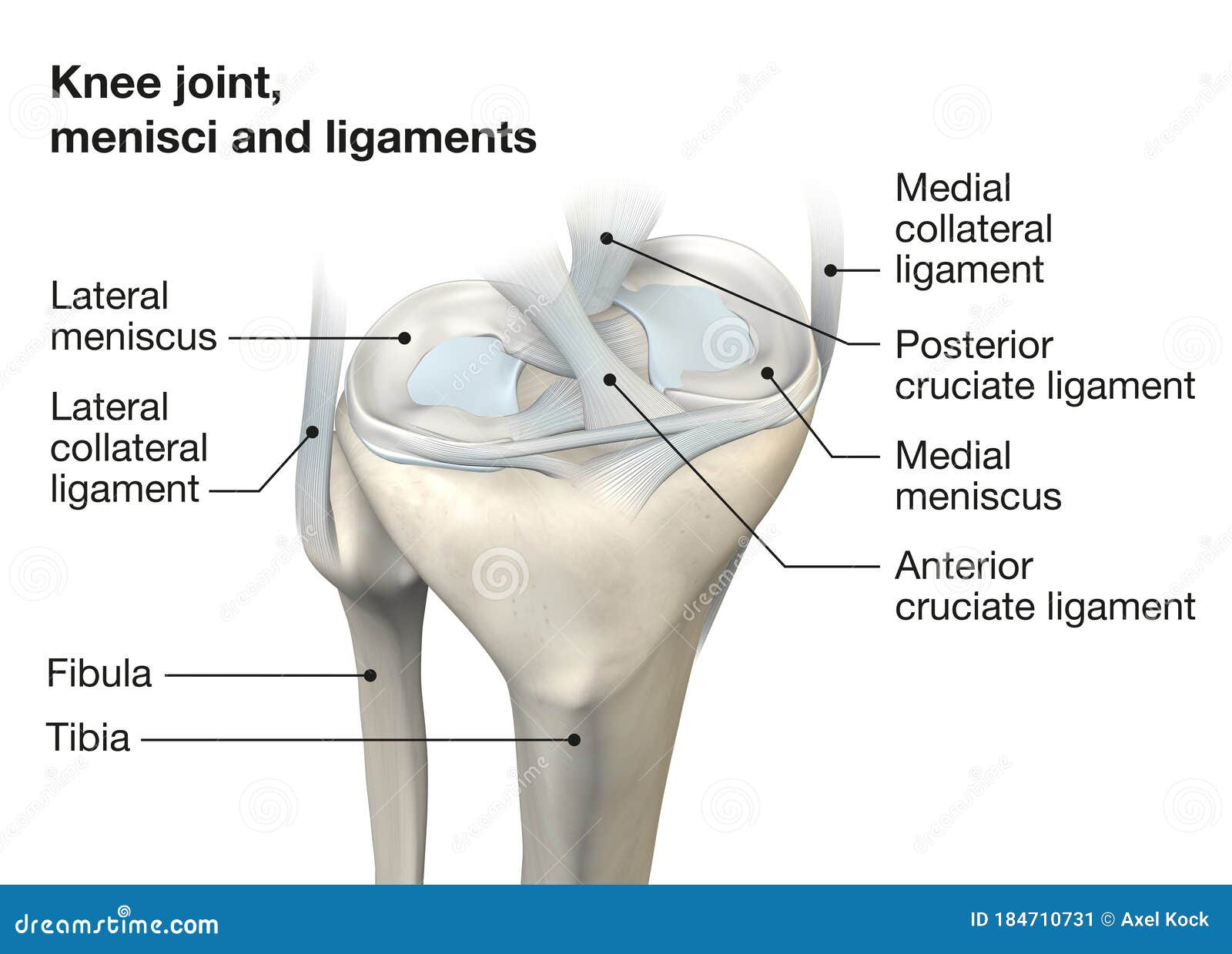
Knee Joint Anatomy, Menisci and Ligaments, Medically 3D Illustration
Knee Joint Anatomy Drawing Colored Digital Download Etsy
Knee Joint Showing Interior Ligaments ClipArt ETC

Structure of the Human Knee Stock Vector Illustration of ligaments
Anatomy of the Knee Joint (With Diagrams and XRay) Owlcation
.jpeg)
Radiopaedia Drawing Bones of the knee joint no labels AnatomyTOOL
Find & Download Free Graphic Resources For Knee Joint Drawing.
It Is A Complex Hinge Joint Composed Of Two Articulations;
Free For Commercial Use High Quality Images
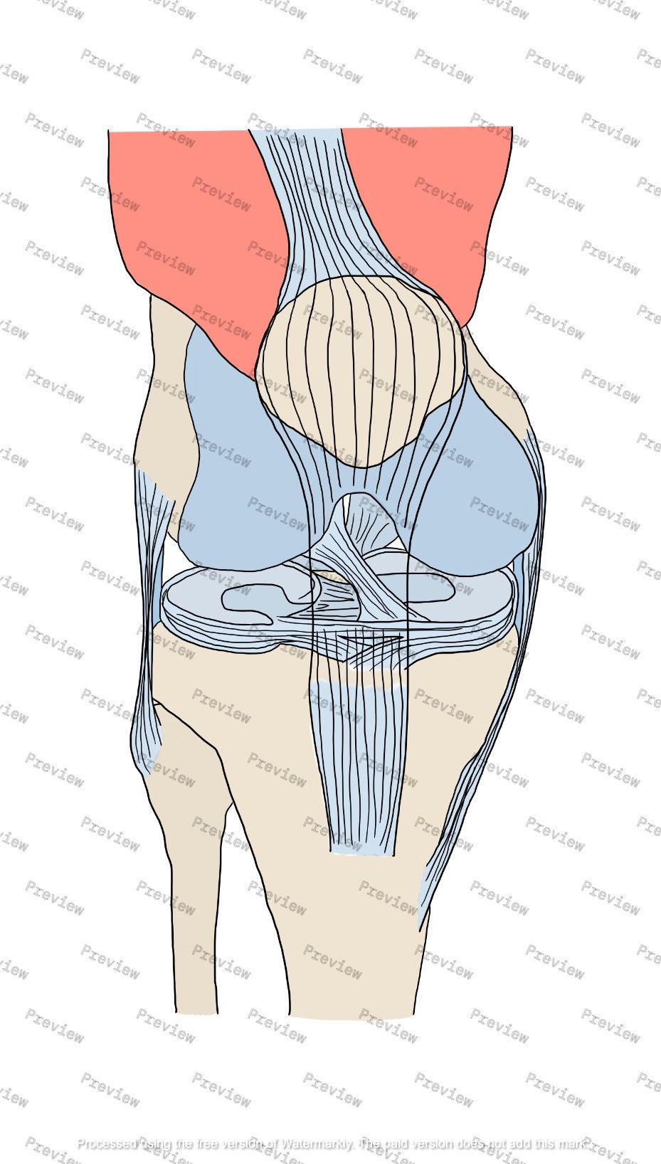
This Image Shows The Knee Joint And Patella From Ventral, Medial And Dorsal.
Related Post: